
Fájl:PBP catalysis.svg

Eredeti fájl (SVG fájl, névlegesen 1 142 × 1 567 képpont, fájlméret: 1,44 MB)
|
Ez a fájl a Wikimedia Commonsból származik. Az alább látható leírás az ottani dokumentációjának másolata. A Commons projekt szabad licencű kép- és multimédiatár. Segíts te is az építésében! |
Összefoglaló
| Leírás |
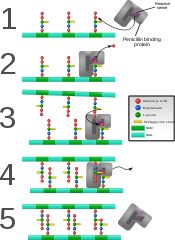
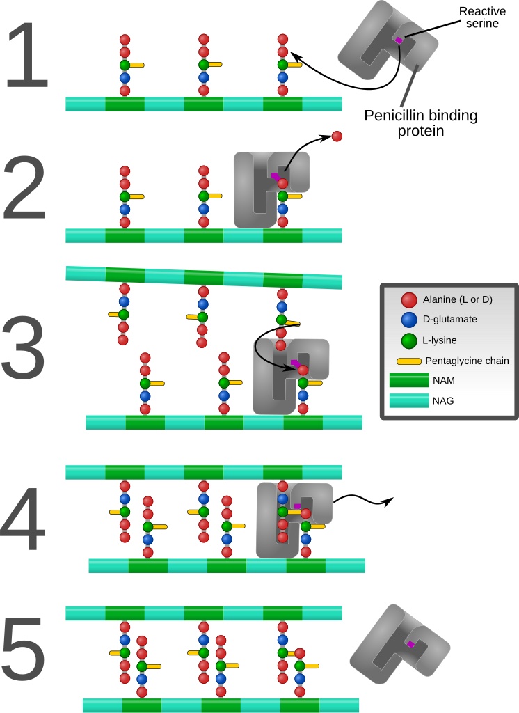
English: Diagram depicting formation of cross-links in the bacterial cell wall by a penicillin binding protein (PBP, an enzyme).
1. The bacterial cell wall consists of strands of repeating N-acetylglucosamine (NAG) and N-acetylmuramic acid (NAM) subunits. The NAM subunits have short peptide chains attached to them. (The exact composition of these can vary. The proximal alanine is usually L-ala and the distal two are usually D-ala.) These chains, in turn, are bound to chains of 5 glycine residues that will be used in cross-linking. 2. The PBP forms a bond with the peptide side chain at the second most distal alanine residue. This displaces the most distal alanine residue. 3. Another strand of bacterial cell wall arrives. The free end of one of the pentaglycine chains displaces the PBP and forms a bond with the terminal alanine on the other strand. 4. After being displaced, the PBP diffuses away. 5. The formation of one cross-link is complete. |
| Dátum | |
| Forrás | A feltöltő saját munkája |
| Szerző | Mcstrother |
| Más változatok |
[]
|


Licenc
- A következőket teheted a művel:
- megoszthatod – szabadon másolhatod, terjesztheted, bemutathatod és előadhatod a művet
- feldolgozhatod – származékos műveket hozhatsz létre
- Az alábbi feltételekkel:
- Nevezd meg! – A szerzőt megfelelően fel kell tüntetned, hivatkozást kell létrehoznod a licencre és jelezned kell, ha a művön változtatást hajtottál végre. Ezt bármilyen észszerű módon megteheted, kivéve oly módon, ami azt sugallná hogy a jogosult támogat téged vagy a felhasználásod körülményeit.
Fájltörténet
Kattints egy időpontra, hogy a fájl akkori állapotát láthasd.
| Dátum/idő | Bélyegkép | Felbontás | Feltöltő | Megjegyzés | |
|---|---|---|---|---|---|
| aktuális | 2011. szeptember 9., 21:26 | | 1 142 × 1 567 (1,44 MB) | Mcstrother | Major revision. Corrected inaccuracies in previous image. |
| 2011. május 3., 06:15 |  | 1 139 × 1 062 (850 KB) | Mcstrother | Changed all fonts to Liberation Sans | |
| 2011. április 10., 05:46 |  | 1 139 × 1 062 (850 KB) | Mcstrother | Changed color of carbohydrate chain. | |
| 2011. március 7., 05:30 |  | 1 139 × 1 062 (835 KB) | Mcstrother | {{Information |Description ={{en|1=Diagram depicting formation of cross-links in the bacterial cell wall by a penicillin binding protein (PBP, an enzyme). 1. The bacterial cell wall consists of strands of repeating N-acetylglucosamine (NAG) and N-ace |
Fájlhasználat
Az alábbi lap használja ezt a fájlt:
Globális fájlhasználat
A következő wikik használják ezt a fájlt:
- Használata itt: en.wikipedia.org
- Használata itt: es.wikipedia.org
- Használata itt: fa.wikipedia.org
- Használata itt: ga.wikipedia.org
- Használata itt: gl.wikipedia.org
- Használata itt: it.wikipedia.org
- Használata itt: mk.wikipedia.org
- Használata itt: th.wikipedia.org