
Fájl:Nematocyst discharge.png
Nem érhető el nagyobb felbontású változat.
Nematocyst_discharge.png (480 × 371 képpont, fájlméret: 190 KB, MIME-típus: image/png)
|
Ez a fájl a Wikimedia Commonsból származik. Az alább látható leírás az ottani dokumentációjának másolata. A Commons projekt szabad licencű kép- és multimédiatár. Segíts te is az építésében! |

|
Ezt a képet el kellene készíteni vektorgrafika használatával SVG fájlként. A formátumnak számos előnye van; lásd a Commons:Media for cleanup lapot a további információkért. Ha a képnek már elérhető SVG-formátumú változata, töltsd fel. Az SVG feltöltése után cseréld le ezt a sablont a következőre: {{vector version available|új kép neve.svg}}.
|
Összefoglaló
| Leírás |
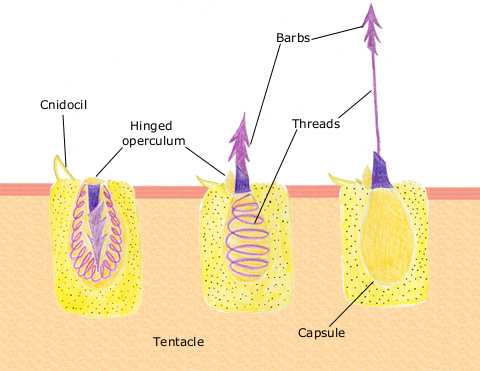
English: The diagram above shows the anatomy of a nematocyst cell and its “firing” sequence, from left to right. On the far left is a nematocyst inside its cellular capsule. The cell’s thread is coiled under pressure and wrapped around a stinging barb. When potential prey makes contact with the tentacles of a polyp, the nematocyst cell is stimulated. This causes a flap of tissue covering the nematocyst—the operculum—to fly open. The middle image shows the open operculum, the rapidly uncoiling thread and the emerging barb. On the far right is the fully extended cell. The barbs at the end of the nematocyst are designed to stick into the polyp’s victim and inject a poisonous liquid. When subdued, the polyp’s tentacles move the prey toward its mouth and the nematocysts recoil back into their capsules. |
| Dátum | 2007. április 11. (eredeti feltöltésének dátuma) |
| Forrás | Áthozva az en.wikipedia projektből a Commonsba. |
| Szerző | Az eredeti feltöltő Spaully a(z) angol Wikipédia projektből volt |
Licenc
Ez a fájl a Creative Commons Így add tovább! 1.0 licenc alapján használható fel
|
Ez a kép közkincs, mert egésze vagy része eredetileg az amerikai Nemzeti Óceán- és Légkörkutatási Hivataltól származik, annak egyik dolgozója készítette munka közben.
|
Eredeti feltöltési napló
Az eredeti leírólap itt volt. Az itt következő felhasználónevek az en.wikipedia projektre hivatkoznak.
- 2007-04-11 17:10 Spaully 480×371×8 (194868 bytes) Modified from: http://www.oceanservice.noaa.gov/education/kits/corals/media/supp_coral01b.html {{Information |Description=Nematocyst discharge process. |Source= Modified from [http://www.oceanservice.noaa.gov/education/kits/corals/media/supp_coral01b.html
Fájltörténet
Kattints egy időpontra, hogy a fájl akkori állapotát láthasd.
| Dátum/idő | Bélyegkép | Felbontás | Feltöltő | Megjegyzés | |
|---|---|---|---|---|---|
| aktuális | 2007. október 13., 19:29 | | 480 × 371 (190 KB) | Alison | {{Information |Description===Description== The diagram above shows the anatomy of a nematocyst cell and its “firing” sequence, from left to right. On the far left is a nematocyst inside its cellular capsule. The cell’s thread is coiled under pressur |
Fájlhasználat
Ezt a fájlt nem használja egyetlen lap sem.
Globális fájlhasználat
A következő wikik használják ezt a fájlt:
- Használata itt: ca.wikipedia.org
- Használata itt: ceb.wikipedia.org
- Használata itt: en.wikipedia.org
- Használata itt: fr.wikipedia.org
- Használata itt: hr.wikipedia.org
- Használata itt: id.wikipedia.org
- Használata itt: it.wikibooks.org
- Használata itt: ja.wikipedia.org
- Használata itt: lv.wikipedia.org
- Használata itt: ms.wikipedia.org
- Használata itt: my.wikipedia.org
- Használata itt: pa.wikipedia.org
- Használata itt: pt.wikipedia.org
- Használata itt: simple.wikipedia.org
- Használata itt: sv.wikipedia.org
- Használata itt: te.wikipedia.org
- Használata itt: th.wikipedia.org
- Használata itt: vi.wikipedia.org